Diagram Of Shoulder And Arm : Arm Definition Bones Muscles Facts Britannica - The shoulder joint is formed where the humerus (upper arm bone) fits into the scapula (shoulder blade), like a ball and.
Diagram Of Shoulder And Arm : Arm Definition Bones Muscles Facts Britannica - The shoulder joint is formed where the humerus (upper arm bone) fits into the scapula (shoulder blade), like a ball and.. This muscle is located near your shoulder. The main shoulder muscles are trapezius, deltoid, pectoralis major and 4 rotator cuff muscles: Where the rounded top of the arm bone (humerus) contacts the shoulder blade is. It allows adduction of your upper arm and flexion of your shoulder. Anatomynote.com found right arm muscle and tendon anatomy from plenty of anatomical pictures on the internet.
Common causes of a shoulder sprain include trauma directly to the shoulder—from a car accident, for example—as well as a fall onto an outstretched arm. A dislocated shoulder occurs when the humerus (upper arm bone) separates from the shoulder blade at the main shoulder joint. Shoulder and arm muscle diagrams. Shoulder joint injuries can be head. It joins with the scapula above at the shoulder joint (or glenohumeral joint) and with the ulna and radius below at the elbow joint.
Muscles Of The Shoulders And Upper Limbs Course Hero from www.coursehero.com The shoulder plays a key role in the blood flow to the arms. The muscles of the shoulder support and produce the movements of the shoulder girdle.they attach the appendicular skeleton of the upper limb to the axial skeleton of the trunk. The shoulder blade is called the scapula and the collarbone is called the clavicle. Common causes of a shoulder sprain include trauma directly to the shoulder—from a car accident, for example—as well as a fall onto an outstretched arm. The shoulder isn't just one bone, it's actually made up of three different bones and various tendons, ligaments, and muscles.the three bones located in the shoulder are the humerus, the scapula, and the clavicle. The humeral head is the ball side. A final test for frozen shoulder is to stand with both arms at the sides and the elbows flexed at ninety degrees. The three bones of the shoulder are the:
What are common rotator cuff injuries?
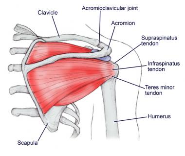
Three of them are located in the anterior compartment — the biceps brachii, brachialis, and coracobrachialis, while the forth is located in the posterior compartment — the triceps brachii). Bones in shoulder, ligaments of the shoulder joint, parts of the shoulder joint, shoulder anatomy, shoulder joints and muscles, shoulder structure anatomy, shoulder tendon anatomy, shoulder tendons ligaments, human muscles, bones in shoulder, ligaments of the shoulder joint, parts of. Where the rounded top of the arm bone (humerus) contacts the shoulder blade is. The main shoulder muscles are trapezius, deltoid, pectoralis major and 4 rotator cuff muscles: There are a number of shoulder bursa located around the joint as shown in the diagram including the: The shoulder isn't just one bone, it's actually made up of three different bones and various tendons, ligaments, and muscles.the three bones located in the shoulder are the humerus, the scapula, and the clavicle. The shoulder girdle includes three bones—the scapula, clavicle and humerus. The shoulder is one of the largest and most complex joints in the body. The bones of the upper arm include the: Shoulder joint injuries can be head. Test your knowledge of the clavicle, scapula and humerus with our labeled diagram exercises and quizzes! Four muscles—the supraspinatus, infraspinatus, teres minor, and subscapularis. This muscle is located near your shoulder.
Shoulder sprains are separated into grades, depending on the extent of damage to the ligaments and the degree of separation between the clavicle and the acromion. It allows adduction of your upper arm and flexion of your shoulder. Diagram of the shoulder, including the location of the rotator cuff. For more anatomy content please follow us and visit our website: The humerus is the (upper) arm bone.
Shoulder Anatomy Best Orthopaedic Doctor For Shoulder Problems Bangalore from bangaloreshoulderinstitute.com The shoulder girdle includes three bones—the scapula, clavicle and humerus. In today's case, our tool of choice is a diagram with the arm muscles clearly labeled. There are a number of shoulder bursa located around the joint as shown in the diagram including the: Subscapularis, supraspinatus, infraspinatus and teres minor. Three of them are located in the anterior compartment — the biceps brachii, brachialis, and coracobrachialis, while the forth is located in the posterior compartment — the triceps brachii). Four muscles—the supraspinatus, infraspinatus, teres minor, and subscapularis. There are four muscles in you upper arm, which is delimited by your shoulder joint and your elbow joint. The list of muscles and their functions are presented below.
We think this is the most useful anatomy picture that you need.
Browse 13,306 shoulder anatomy stock photos and images available, or search for shoulder joint or rotator cuff to find more great stock photos and pictures. The humeral head is the ball side. The main shoulder muscles are trapezius, deltoid, pectoralis major and 4 rotator cuff muscles: This is where the humerus (arm bone) meets the scapula (shoulder blade). Three of them are located in the anterior compartment — the biceps brachii, brachialis, and coracobrachialis, while the forth is located in the posterior compartment — the triceps brachii). This muscle is located near your shoulder. The shoulder plays a key role in the blood flow to the arms. The shoulder is not a single joint, but a complex arrangement of bones, ligaments, muscles, and tendons that is better called the shoulder girdle. We hope this picture right arm muscle and tendon anatomy can help you study and research. The shoulder blade is called the scapula and the collarbone is called the clavicle. Common causes of a shoulder sprain include trauma directly to the shoulder—from a car accident, for example—as well as a fall onto an outstretched arm. The rotator cuff muscles are important stabilizers and movers of the shoulder joint. The upper arm includes the shoulder as well as the area between the shoulder and elbow joint.
The humerus is the bone of the arm that articulates with the scapula proximally and with the radius and the ulna distally. Illustrated representation of the structure and musculature of the human arm. As the name implies, the rotator cuff functions to allow you to rotate your shoulder and lift your arm. The shoulder joint is formed where the humerus (upper arm bone) fits into the scapula (shoulder blade), like a ball and. What are common rotator cuff injuries?
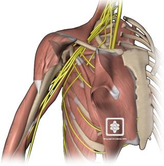
Nerves Of The Shoulder Shoulderdoc from www.shoulderdoc.co.uk A final test for frozen shoulder is to stand with both arms at the sides and the elbows flexed at ninety degrees. There are two joints within the shoulder that can be affected by osteoarthritis. Diagram of the shoulder, including the location of the rotator cuff. As the name implies, the rotator cuff functions to allow you to rotate your shoulder and lift your arm. Where the rounded top of the arm bone (humerus) contacts the shoulder blade is. The largest bone of the arm, the humerus connects to the scapula and clavicle in the shoulder. There are a number of shoulder bursa located around the joint as shown in the diagram including the: This is called the glenoid.
Four of them are found on the anterior aspect of the shoulder, whereas the rest are located on the shoulder's posterior aspect and in the back.
Muscles of the shoulder : Three of them are located in the anterior compartment — the biceps brachii, brachialis, and coracobrachialis, while the forth is located in the posterior compartment — the triceps brachii). This holds your upper arm bone to your shoulder blade and helps you rotate your arm, hold it straight out and lower it. Is the wear and tear of shoulder cartilage until bare bone is exposed. The main shoulder muscles are trapezius, deltoid, pectoralis major and 4 rotator cuff muscles: Anatomynote.com found right arm muscle and tendon anatomy from plenty of anatomical pictures on the internet. The glenoid is covered with smooth cartilage. We think this is the most useful anatomy picture that you need. The shoulder is one of the largest and most complex joints in the body. Located superior to the shoulder joint, the deltoid muscle works with the supraspinatus to abduct the arm at the shoulder. The acromioclavicular joint is formed by an articulation between the lateral end of the clavicle and the acromion process of the scapula. Four of them are found on the anterior aspect of the shoulder, whereas the rest are located on the shoulder's posterior aspect and in the back. The upper arm includes the shoulder as well as the area between the shoulder and elbow joint.
Shoulder muscles move the shoulder blades and upper arm bones diagram of shoulder. The acromioclavicular joint is formed by an articulation between the lateral end of the clavicle and the acromion process of the scapula.


0 Komentar